细胞衰老(Cell senescence)是细胞控制其生长潜能的保障机制,一般含义是复制衰老(Replicative senescence,RS),指正常细胞经过有限次数的分裂后,停止分裂,此时细胞虽然是存活的,但是细胞形态和生理代谢活性发生明显变化的现象。体外衰老细胞研究常用的生物学特征包括:1)不可逆的生长停滞;2)与衰老相关的β-半乳糖苷酶(senescence associated β-galactosidase, SA-β-gal)的活化。作为溶酶体内的水解酶,通常在pH 4.0的条件下表现活性,但是衰老细胞内其在pH 6.0的条件下表现活性,且随着传代次数的增加,细胞群体中表现衰老相关的β-半乳糖苷酶的细胞数目逐渐增加。
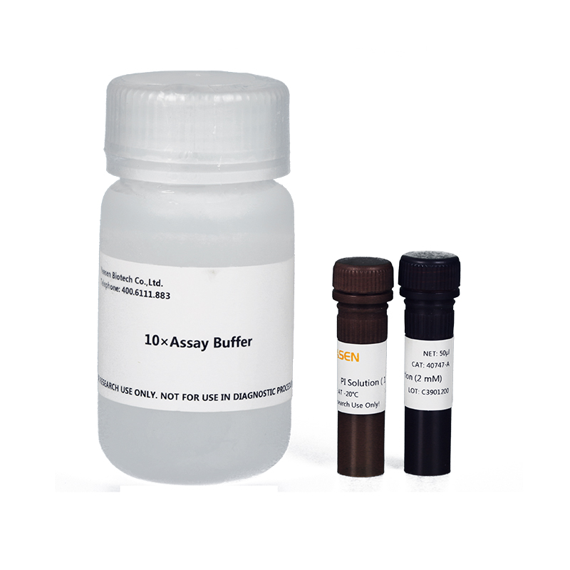
本试剂盒正是基于SA -β-gal活性变化的生物学特征而设计的,以X-Gal为底物,在衰老特异性的β-半乳糖苷酶催化下生成深蓝色产物,从而在光学显微镜下观察颜色变化以分析细胞的衰老状况。
本试剂盒可以培养细胞的衰老检测,也可以用于组织切片的衰老检测。产品性能稳定,参数经优化,着色敏感,特异性高。仅对衰老细胞进行染色,不会染色衰老前的细胞(presenescent cells)、静止期细胞(quiescent cells)、永生细胞(immortal cells)或肿瘤细胞。
冰袋运输。-20℃保存,一年有效,其中X-Gal溶液需避光保存。
Q:细胞衰老β-半乳糖苷酶染色试剂盒能否用于石蜡切片?
A:在石蜡包埋的过程中,温度、固定液等因素可能会导致β-半乳糖苷酶失活,从而造成染色失败,因此本试剂盒不建议用于石蜡切片的衰老检测。
Q:着色很浅或未染上色,可能是什么原因?
A:1.样本无衰老情况:如果染色的样本没有诱导成功或衰老程度不明显,从而导致染色的时候着色较浅或者未染上色。
2.试剂未完全溶解:β-半乳糖苷酶染色液解冻后若有沉淀,必须完全溶解无沉淀之后才可以使用。
3.冰冻切片放置过久:冰冻切片时间较长,β-半乳糖苷酶可能降解了。
4.染色时可能把样本放在细胞培养箱进行染色:细胞衰老β-半乳糖苷酶染色反应依赖于特定的pH条件,不能在二氧化碳培养箱中进行染色反应,用于细胞培养的二氧化碳培养箱中较高浓度的二氧化碳会影响染色工作液的pH值,而导致染色失败。
[1] Zhang D, Liu Y, Zhu Y, et al. A non-canonical cGAS-STING-PERK pathway facilitates the translational program critical for senescence and organ fibrosis. Nat Cell Biol. 2022;24(5):766-782. doi:10.1038/s41556-022-00894-z(IF:28.824)
[2] Meng F, Yu Z, Zhang D, et al. Induced phase separation of mutant NF2 imprisons the cGAS-STING machinery to abrogate antitumor immunity. Mol Cell. 2021;81(20):4147-4164.e7. doi:10.1016/j.molcel.2021.07.040(IF:17.970)
[3] Yang Y, Liu S, He C, et al. LncRNA LYPLAL1-AS1 rejuvenates human adipose-derived mesenchymal stem cell senescence via transcriptional MIRLET7B inactivation. Cell Biosci. 2022;12(1):45. Published 2022 Apr 21. doi:10.1186/s13578-022-00782-x(IF:7.133)
[4] Adili A, Zhu X, Cao H, et al. Atrial Fibrillation Underlies Cardiomyocyte Senescence and Contributes to Deleterious Atrial Remodeling during Disease Progression. Aging Dis. 2022;13(1):298-312. Published 2022 Feb 1. doi:10.14336/AD.2021.0619(IF:6.745)
[5] Qing Y, Li H, Zhao Y, et al. One-Two Punch Therapy for the Treatment of T-Cell Malignancies Involving p53-Dependent Cellular Senescence. Oxid Med Cell Longev. 2021;2021:5529518. Published 2021 Sep 21. doi:10.1155/2021/5529518(IF:6.543)
[6] Gu W, Wang L, Gu R, et al. Defects of cohesin loader lead to bone dysplasia associated with transcriptional disturbance. J Cell Physiol. 2021;236(12):8208-8225. doi:10.1002/jcp.30491(IF:6.384)
[7] Xie L, Huang W, Fang Z, et al. CircERCC2 ameliorated intervertebral disc degeneration by regulating mitophagy and apoptosis through miR-182-5p/SIRT1 axis. Cell Death Dis. 2019;10(10):751. Published 2019 Oct 3. doi:10.1038/s41419-019-1978-2(IF:5.959)
[8] Yang D, Guo Q, Liang Y, et al. Wogonin induces cellular senescence in breast cancer via suppressing TXNRD2 expression. Arch Toxicol. 2020;94(10):3433-3447. doi:10.1007/s00204-020-02842-y(IF:5.059)
[9] Fan J, An X, Yang Y, et al. MiR-1292 Targets FZD4 to Regulate Senescence and Osteogenic Differentiation of Stem Cells in TE/SJ/Mesenchymal Tissue System via the Wnt/β-catenin Pathway. Aging Dis. 2018;9(6):1103-1121. Published 2018 Dec 4. doi:10.14336/AD.2018.1110(IF:5.058)
[10] Yang D, Tian X, Ye Y, et al. Identification of GL-V9 as a novel senolytic agent against senescent breast cancer cells. Life Sci. 2021;272:119196. doi:10.1016/j.lfs.2021.119196(IF:5.037)
[11] Wang J, Wang WN, Xu SB, et al. MicroRNA-214-3p: A link between autophagy and endothelial cell dysfunction in atherosclerosis. Acta Physiol (Oxf). 2018;222(3):10.1111/apha.12973. doi:10.1111/apha.12973(IF:4.867)
[12] Wang D, Cai G, Wang H, He J. TRAF3, a Target of MicroRNA-363-3p, Suppresses Senescence and Regulates the Balance Between Osteoblastic and Adipocytic Differentiation of Rat Bone Marrow-Derived Mesenchymal Stem Cells. Stem Cells Dev. 2020;29(11):737-745. doi:10.1089/scd.2019.0276(IF:3.153)


